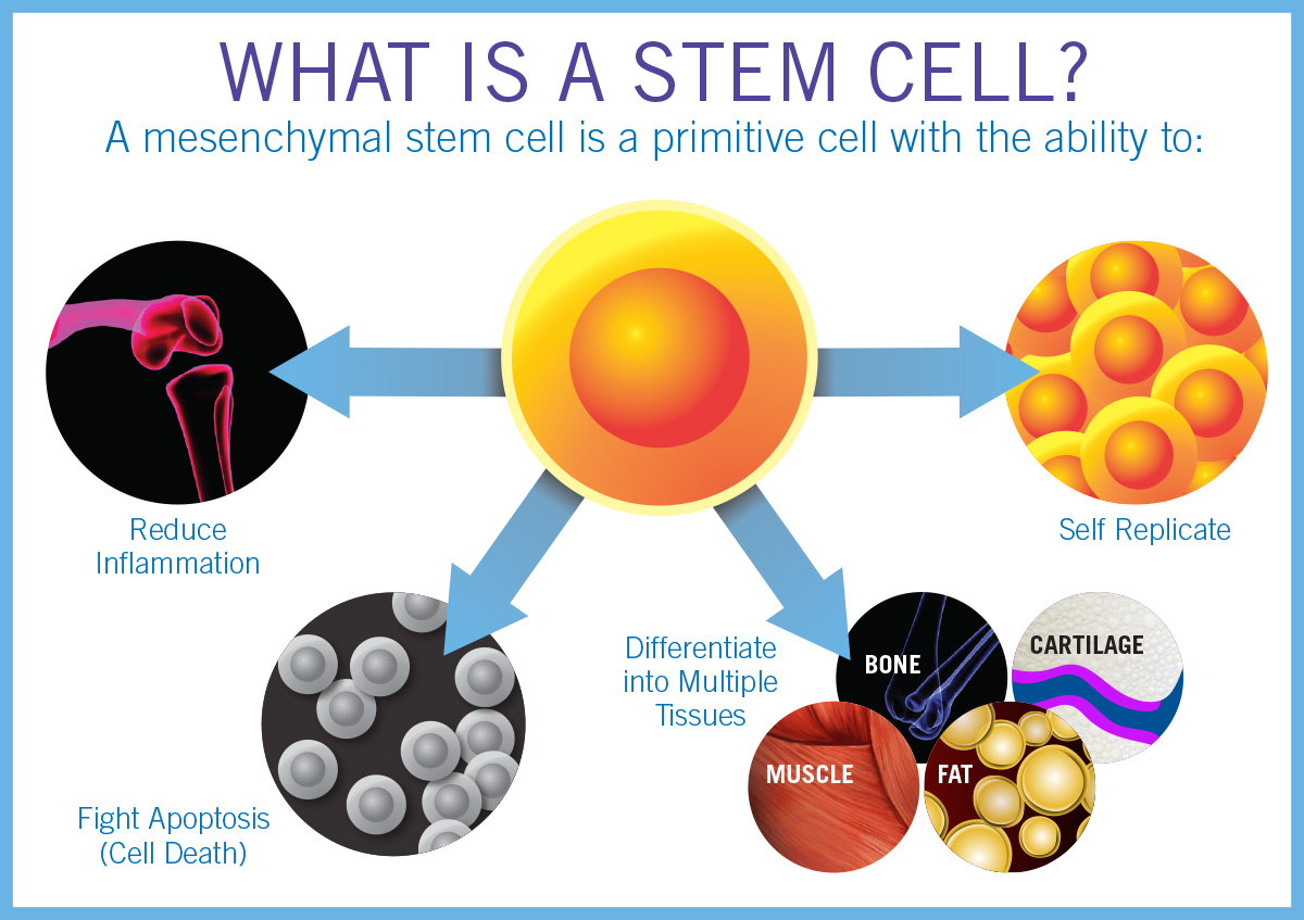
Stem cell therapy is revolutionizing the approach to treating eye damage, bringing new hope to patients with conditions once deemed untreatable. Recent clinical trials at Mass Eye and Ear have demonstrated the efficacy of this innovative technique, specifically focusing on cultivated autologous limbal epithelial cells (CALEC) for restoring damaged corneal surfaces. By utilizing stem cells harvested from healthy eyes, researchers are facilitating significant advancements in eye damage treatment, offering a viable alternative to traditional transplantation methods. This breakthrough could significantly improve the quality of life for individuals suffering from chronic eye injuries, with promising results aiming to enhance corneal surface restoration. As ocular stem cell research progresses, the implications for therapy using limbal epithelial cells may become vital in combating a range of vision-threatening issues.
Revolutionary advancements in ocular regenerative medicine are exemplified by the innovative application of stem cell-based treatment for ocular injuries. These exciting developments tap into the potential of cultivating specific eye cells to repair corneal damage effectively. Known as cultivated autologous limbal epithelial cells (CALEC), this technique has emerged as a beacon of hope for those experiencing severe vision impairment due to corneal surface degradation. By harnessing the body’s innate healing properties through the proliferation of stem cells, researchers are paving the way for new protocols that restore sight and comfort to patients. This approach not only signifies progress in eye damage treatment but also underscores the importance of continuous research in ocular cell therapy.
Overview of the CALEC Procedure
The Cultivated Autologous Limbal Epithelial Cells (CALEC) procedure is an innovative treatment designed to address severe corneal damage that previously had no effective remedy. This procedure aims to restore the cornea’s surface using the patient’s own stem cells harvested from a healthy eye. The technique involves a meticulous process that begins with obtaining limbal epithelial cells through a biopsy. After careful cultivation in a lab, these cells are transformed into a tissue graft that can be transplanted onto the affected cornea. The groundbreaking work conducted at Mass Eye and Ear has already showcased promising results, with over 90% of patients experiencing significant improvements in corneal restoration within an 18-month follow-up period.
The success of the CALEC procedure not only signifies a leap forward in ocular stem cell research but also offers hope for individuals facing chronic vision issues due to corneal injuries. By targeting injuries that cannot be treated by traditional corneal transplant methods, the CALEC procedure exemplifies how scientific advancement can address previously untreatable conditions. It restores functionality in patients’ eyes and alleviates related symptoms such as chronic pain and discomfort.
In clinical settings, investigators emphasize the importance of having a reliable supply of limbal epithelial cells for the CALEC procedure. Currently, this method is limited to patients with only one damaged eye, which makes the feasibility of broader applications a subject of ongoing research. Plans are in the works to develop an allogeneic manufacturing process. This approach aims to source limbal stem cells from healthy cadaveric donor eyes, thereby expanding the treatment options for patients with bilateral corneal damage. As research continues, the eyes of many patients rest on the potential of CALEC to redefine the landscape of eye damage treatment and significantly enhance their quality of life.
The Impact of Ocular Stem Cell Research on Eye Damage Treatment
Ocular stem cell research has emerged as a vital frontier in the quest for advanced eye damage treatment modalities. The CALEC procedure is a prime example of how this field has begun to make significant strides in offering solutions to conditions previously deemed irreversible. Researchers at Mass Eye and Ear have pioneered methods to cultivate limbal epithelial cells efficiently, creating viable grafts for patients suffering from chronic corneal damage. This innovation symbolizes a shift towards personalized regenerative therapies where treatment is tailored to each patient’s specific condition, rather than relying solely on donor tissues.
In addition to the immediate benefits that come from effective restoration of the corneal surface, the implications of such advancements can radically alter the future of ocular health. The successful incorporation of stem cell therapy into clinical practice not only facilitates healing but also heralds a future where novel protocols could reduce the incidence of blindness associated with corneal injuries. The ongoing investigations will likely unravel more possibilities, leading to wider applications of stem cell treatments across various ocular conditions.
As eye injury prevalence continues to rise due to environmental factors and non-communicable diseases, researchers are increasingly focusing on maximizing the potential of stem cells in eye health. Through studies like the CALEC trial, empirical data highlights the success rates and safety profiles of stem cell therapies, attracting attention from funding bodies like the National Eye Institute. This increased interest accelerates funding toward ocular stem cell research, which, in turn, may expedite the development of treatments for patients suffering from ocular diseases or injuries. The pathway paved by CALEC shows promise not only for restoring vision but also for contributing to the overall understanding of ocular biology, opening doors to revolutionary therapies.
Future Directions in Corneal Stem Cell Treatments
The future of corneal stem cell treatments looks hopeful, particularly with advancements in the CALEC procedure and ongoing research into ocular stem cells. As the field evolves, researchers strive for larger-scale clinical trials, which would provide more data on the efficacy and safety of therapies derived from limbal stem cells. Initiatives focusing on multi-center studies may help validate the outcomes observed in smaller populations while also addressing issues like long-term sustainability of results and the necessary regulatory processes for wider implementation.
The potential for an allogeneic process also looms large in the future, which could allow patients with bilateral eye injuries to benefit from stem cell therapies. Such a breakthrough would significantly multiply the number of individuals eligible for treatment, enhancing the quality of life for countless patients suffering from persistent corneal damage.
Researchers are also investigating the integration of cutting-edge technologies like tissue engineering and gene therapy alongside traditional stem cell approaches. This merging of disciplines promises to further revolutionize ocular treatments by not only repairing damage but also renewing cell health and function at a molecular level. Future studies are likely to investigate combinations that optimize the healing process, assess the durability of results over extended periods, and evaluate the impact on patients’ overall ocular health. By building on the successes and lessons learned from the CALEC trial, the field of ocular stem cell research is poised to reshape the landscape of eye care profoundly.
Understanding Corneal Surface Restoration
Corneal surface restoration is a critical aspect of vision health that significantly impacts an individual’s quality of life. The cornea’s smooth surface is essential for proper light refraction, clarity of vision, and protection from external elements. When this surface is compromised due to injuries such as chemical burns, infections, or trauma, patients can experience severe vision impairment and chronic discomfort. Restoration methods, like the recently developed CALEC procedure, emphasize the need to not just address the symptoms but to fundamentally restore the cornea’s structural integrity using biological means.
Corneal surface restoration technologies often employ the principles of regenerative medicine, which encourages the body’s intrinsic healing capabilities through various methodologies. The CALEC procedure utilizes a graft made from cultivated limbal epithelial cells, effectively providing the cornea with the necessary components for recovery. The success of this treatment has possibilities extending beyond those previously felt impossible, allowing patients with extensive corneal damage to regain significant functional vision.
Various factors contribute to the effectiveness of corneal surface restoration techniques, such as the initial health of the limbal epithelial cells, the precision of the surgical procedure, and the postoperative care provided to patients. Emerging research continuously seeks to enhance these techniques, striving for faster recovery times, reduced risks of complications, and improved overall outcomes. Studies exploring the impact of adjunct therapies, including anti-inflammatory medications and innovative post-surgical support, demonstrate a holistic approach focusing on both the surgical process and subsequent care to optimize patient recovery. As the science behind corneal surface restoration advances, it holds the promise of transforming how individuals with eye damage are treated today.
Challenges in Limbal Epithelial Cell Harvesting
Harvesting limbal epithelial cells presents several challenges that impact the effectiveness of regenerative therapies like the CALEC procedure. Primarily, the technique requires that patients have healthy limbal epithelial cells in at least one eye, which limits the candidate pool. Those suffering from bilateral corneal damage cannot utilize this method unless alternative strategies are developed. Effective harvesting requires precision and planning, as any damage to the healthy eye during the biosampling process could lead to further complications or vision problems.
Additionally, the cultivation process must adhere to stringent regulatory standards as established by health authorities, ensuring safety and efficacy in the produced grafts. The complexity of managing cell cultures and ensuring viable cell multiplication requires specialized facilities that may not be readily available across all health institutions.
The reliance on a donor eye for extracting limbal epithelial cells also raises ethical questions pertaining to consent and availability, particularly for postmortem retrieval. As ocular stem cell research progresses, it is crucial to address these challenges to proficiently expand the eligibility of patients needing corneal restoration. Developing methodologies that lessen the reliance on patient-specific cells may optimize outcomes for many individuals suffering from various degrees of corneal injuries. This may include leveraging advances in tissue engineering or identifying alternative stem cell sources. Addressing these challenges head-on will only enhance the field’s ability to offer effective and accessible ocular treatments.
The Role of FDA Approval in Stem Cell Treatments
Obtaining U.S. Food and Drug Administration (FDA) approval is a pivotal step for any new therapeutic intervention, especially in the field of stem cell treatments for eye damage. The rigorous evaluation ensures that new methodologies like the CALEC procedure not only meet safety benchmarks but also demonstrate consistent efficacy across diverse patient populations. As the first human study funded by the National Eye Institute focusing on stem cell therapy for eye conditions, the success of the CALEC clinical trial will serve as a benchmark for future treatments seeking FDA endorsement.
The approval process involves extensive data collection and analysis, highlighting patient outcomes, safety profiles, and the overall applicability of treatment across various demographics. This transparency builds trust among stakeholders, including patients, healthcare professionals, and potential investors, in the efficacy of pioneering therapies. Furthermore, engaging in dialogue with the FDA early in the development process can significantly streamline the path to approval for innovative treatments.
Ensuring that new treatments adhere to established safety regulations and deliver verifiable benefits to patients is fundamental to fostering a responsible and ethical approach in the realm of ocular regenerative medicine. As the CALEC procedure progresses toward broader application, collaboration between researchers and regulatory bodies will play a crucial role in establishing guidelines and protocols necessary for future stem cell therapies. This process not only impacts the CALEC approach but also shapes the regulatory landscape as a whole, guiding forthcoming innovations in ocular health.
Potential Benefits of the CALEC Procedure
The potential benefits of the CALEC procedure extend beyond mere restoration of vision; they encompass the improvement of overall quality of life for individuals suffering from corneal damage. With a success rate reported at over 90%, the CALEC procedure represents a beacon of hope for those who have endured chronic pain and suffering due to their eye injuries. Not only does this treatment help reinstate proper corneal function, but it also significantly reduces the debilitating symptoms associated with untreated corneal damage, such as constant discomfort and visual impairment.
Furthermore, the innovative nature of the CALEC procedure enhances the versatility of treatment options available in ophthalmology. By cultivating the patient’s own cells, this method minimizes the risk of rejection or other complications that are often associated with donor grafts. This personalized approach not only leads to more favorable outcomes post-surgery but also positions the CALEC procedure as a model for future regenerative therapies across various medical fields.
Additionally, as patient outcomes from the CALEC procedure are documented and analyzed over time, the knowledge gained can inform best practices for managing corneal injuries. Researchers aim to collect extensive data on complications, recovery times, and visual acuity improvements, further refining the approach and potentially establishing standardized protocols that can be implemented across various healthcare settings. As the field of ocular stem cell research continues to grow, the lessons learned from CALEC might contribute to more advanced therapies and methodologies that enhance the overall effectiveness of treatments for eye damage.
Frequently Asked Questions
What is stem cell therapy and how does it relate to eye damage treatment?
Stem cell therapy involves using specialized cells that can develop into different types of cells in the body. In the context of eye damage treatment, specifically for corneal injuries, stem cell therapy employs cultivated autologous limbal epithelial cells (CALEC). This innovative approach helps restore the cornea’s surface by using stem cells harvested from a healthy eye to regenerate the damaged areas of the cornea.
How effective is the CALEC procedure for restoring corneal surfaces?
The CALEC procedure has demonstrated over 90 percent effectiveness in restoring corneal surfaces in clinical trials. Within just three months, 50 percent of the patients had their corneas fully restored, with success rates rising to 79 percent at twelve months and 77 percent at eighteen months post-treatment.
What role do limbal epithelial cells play in corneal health?
Limbal epithelial cells, harvested from the limbus of the eye, are crucial for maintaining the smoothness and health of the cornea. They help replenish the corneal surface when it is damaged. A deficiency in these cells can result from injuries like chemical burns or infections, leading to chronic damage and pain without the possibility of corneal transplant.
What steps are involved in the CALEC procedure for ocular stem cell research?
The CALEC procedure begins with a biopsy from the healthy eye to obtain limbal epithelial cells. These cells are then cultivated into a tissue graft through a specialized process which takes two to three weeks. After that, the graft is surgically transplanted into the damaged eye to restore its surface and function.
Is the CALEC procedure currently available for patients with corneal damage?
As of now, the CALEC procedure remains experimental and is not widely available in U.S. hospitals. Additional research and trials are necessary before seeking FDA approval to make this treatment more accessible for patients suffering from corneal damage.
What potential does the CALEC procedure have for patients with bilateral eye injuries?
Currently, the CALEC procedure can only be performed on patients with one affected eye, as it requires harvesting cells from a healthy eye. However, researchers aim to develop an allogeneic process using limbal stem cells from cadaveric donor eyes, which would expand treatment options for patients with damage in both eyes.
What safety profile does the CALEC procedure show in clinical trials?
Clinical trials for the CALEC procedure indicate a high safety profile, with no severe complications reported in donor or recipient eyes. Though there was one case of a bacterial infection, it was linked to external factors rather than the procedure itself, indicating its overall safety for patients.
How does CALEC compare to traditional methods for treating severe corneal injuries?
Traditional methods, like corneal transplants, may not be viable options for patients with severe, chronic corneal injuries due to limbal stem cell deficiency. The CALEC procedure offers a novel solution by regenerating the corneal surface using the patient’s own stem cells, potentially restoring vision where conventional strategies have failed.
What are the future prospects for stem cell therapy in ocular treatments?
The future of stem cell therapy in ocular treatments looks promising, especially with ongoing research into the CALEC procedure. Scientists hope to initiate further clinical trials to confirm efficacy across larger patient groups, shorten manufacturing times, and work towards FDA approval, potentially transforming treatment for various eye conditions.
What organizations are involved in the development of stem cell therapies for eye diseases?
The development of stem cell therapies for eye diseases involves collaboration between reputable institutions such as Mass Eye and Ear, Dana-Farber Cancer Institute, and Boston Children’s Hospital. These partnerships along with funding from the National Eye Institute highlight the commitment to advancing ocular stem cell research and therapies.
| Key Points |
|---|
| Ula Jurkunas leads the first CALEC surgery at Mass Eye and Ear. |
| Stem cell therapy is used to repair damaged corneal surfaces in clinical trials. |
| Successful outcomes for 14 patients over 18 months, with a 90% effectiveness rate. |
| The CALEC procedure involves harvesting stem cells from a healthy eye. |
| Potential benefits for patients with chronic corneal damage previously deemed untreatable. |
| Future research aims to expand the application of CALEC to patients with damage in both eyes. |
Summary
Stem cell therapy offers groundbreaking solutions for individuals suffering from severe corneal damage. Recent trials at Mass Eye and Ear have demonstrated that cultivated autologous limbal epithelial cells (CALEC), harvested from healthy eyes, can effectively restore damaged corneal surfaces, showing a remarkable success rate of over 93%. As we look towards the future, continued advancements in stem cell therapy promise not only to enhance recovery rates but also to broaden treatment accessibility, potentially transforming the landscape of ophthalmology and restoring hope for many.
